


Description
Key Features:
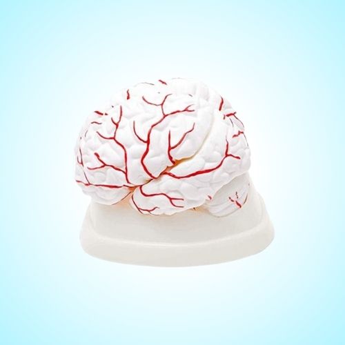
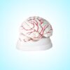
Realistic External Brain Anatomy
-
Cerebral Hemisphere: Shows central sulcus, lateral and parieto-occipital fissures, and clearly labeled lobes—frontal, parietal, temporal, and occipital.
-
Median & Basal Surfaces: Displays key elements such as the corpus callosum (cross-section), calcarine fissure, olfactory bulb and tract.
Detailed Brain Stem
-
Dorsal View: Includes the thalamus, pulvinar, corpora quadrigemina, and rhomboid fossa.
-
Ventral View: Shows the optic chiasma, mammillary bodies, crura pedunculi, and pons.
Cerebellum Clarity
-
Features distinct cerebellar hemispheres and vermis to help identify structural components with ease.
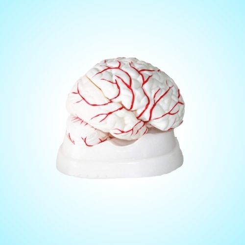
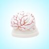
Comprehensive Arterial Supply
-
Supplied by: Two vertebral and two internal carotid arteries.
-
Basilar Artery: Formed by vertebral arteries, branching into superior cerebellar and posterior cerebral arteries.
-
Cerebral Arteries: Includes posterior, middle, and anterior cerebral arteries with interconnecting branches.
-
Cerebellar Arteries: Three pairs clearly represented—posterior inferior, anterior inferior, and superior cerebellar arteries.
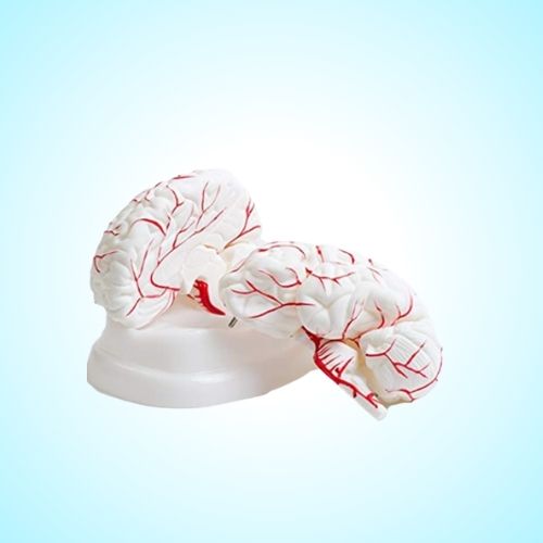
Construction & Usability
-
Material: Made of durable, high-quality plastic.
-
Dissectible: 8-piece detachable design, mounted on a sturdy base for easy reassembly and thorough exploration.
-
Size: Life-size for realistic spatial understanding and demonstration.
Perfect For:
-
Medical and paramedical students studying anatomy and physiology
-
Educators and institutions teaching brain structure and function
-
Healthcare professionals requiring a reliable visual reference
Reviews
There are no reviews yet.